The Centers for Advanced Orthopaedics is redefining the way musculoskeletal care is delivered across the region with locations throughout Maryland, DC, Virginia and Pennsylvania.
4 Different Types Of Imaging Tests For Foot Conditions

Orthopedic specialists have a number of tools in their arsenal to help pinpoint exactly what’s going on in a person’s foot and ankle. These imaging tests provide a much clearer picture of the inner workings of the foot, and different tests can showcase different structures in the area. In today’s blog, we’re going to take a closer look at four different kinds of imaging tests commonly used to help diagnose foot and ankle issues, and we’ll explain which conditions they often help discover.
Four Different Types Of Imaging Tests
If you’re dealing with foot or ankle pain and it’s not obvious what’s going on, your orthopedic specialist may recommend you undergo one of these four imaging tests:
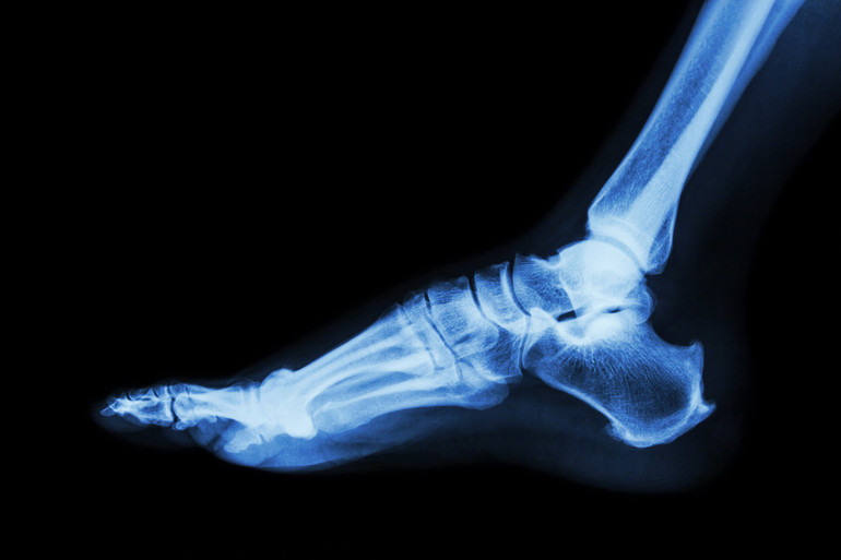
- X-Ray - The x-ray (radiograph) is the most common and widely available diagnostic imaging technique, and it’s especially helpful in identifying issues with bones. A X-ray relies on electromagnetic waves to produce an image that showcases bones, their density and any fractures or deposits that have formed in the area. It is the least detailed of the four techniques we’ll review, but it is also the cheapest and most widely available at clinics, and it can be just what the patient needs if they are dealing with a problem like a fracture or bunion. X-rays are also helpful after a procedure to ensure bone healing is going as expected or to ensure hardware hasn’t shifted.
- CT Scan - CT scan stands for computer tomography, and it’s a more advanced form of an X-ray. With this technique, X-rays are combined with computer technology to create a more detailed, cross-sectional image of your foot. This allows your doctor to view the issue on multiple planes to understand the true nature of the issue. Doctors can see the size, shape and position of issues within the foot, making it helpful for diagnosing conditions like arthritis, fractures, infections, tumors and other deformities.
- MRI - An MRI is an even more detailed imaging technique that doesn’t involve radiation like X-rays or CT scans. Instead, it employs a large magnet and radio waves to produce a three-dimensional image of your foot. It also helps to highlight both bone and soft tissues like tendons or ligaments, making it helpful for diagnosing a wide range of conditions. Not only can it identify problems like arthritis, fractures, infections and tumors, but it can also pinpoint tears or damage to tendons, ligaments and cartilage. An MRI takes more time to complete than the two above techniques, and because a large magnet is used, it may not be appropriate for people with certain implants or cardiac devices.
- Ultrasound - You may be familiar with ultrasound technology if you’ve ever had children, but the technique is also helpful for spotting certain foot and ankle conditions. Ultrasound uses targeted sound waves, which are then bounced back to a recording device to produce an image. It’s a safe, noninvasive and painless diagnostic procedure, and it’s great for identifying certain conditions like tarsal tunnel syndrome, tendonitis, plantar fasciitis, bursitis, Morton’s neuroma or for injuries to tendons, ligaments and cartilage.
So the next time you head to the doctor’s office with a foot or ankle injury, don’t be surprised if they turn to one of these imaging techniques to provide a precise diagnosis. We have all these technologies at our disposal, so if you truly want to understand what’s going on in your foot or ankle, reach out to the team at The Centers For Advanced Orthopaedics today.